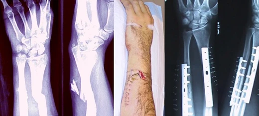
Colles' fracture
Irish surgeon who described the distal radius fracture in 1814.

A Colles' fracture is a break near the wrist in the larger bone of the forearm, the radius, where the broken end tilts backward. People with this injury often experience pain, swelling, a visible deformity, and bruising. A possible complication is injury to the median nerve. The usual cause is falling onto an outstretched hand. Having osteoporosis raises the risk. Doctors typically confirm the fracture with X-rays. The small bone on the pinky side of the forearm, the ulna, may also be fractured at its tip. Treatment options include wearing a cast or undergoing surgery. About 15 percent of individuals will experience a Colles' fracture at some point in their lives. It is more common in young adults and older people than in children or middle-aged adults, and women are affected more often than men. The injury is named after the Irish surgeon Abraham Colles, who first described it in 1814.
- born
- 1773
- died
- 1843
- field
- Surgery
- nationality
- Irish
- known_for
- Describing the Colles' fracture in 1814
Lore & Background
Abraham Colles (1773–1843) was an Irish surgeon from Kilkenny. He first described the fracture that bears his name in 1814, before the advent of X-rays, by simply looking at the classic deformity. The fracture is sometimes referred to as a 'dinner fork' or 'bayonet' deformity due to the shape of the resultant forearm. It is sometimes said that Claude Pouteau was the first to describe the Colles' fracture (which is sometimes called the Pouteau-Colles fracture), but, according to P. Liverneaux, it is not the case.
Reader's Guide
The Colles' fracture remains a common clinical entity, occurring in about 15% of people at some point in their lives. It occurs more commonly in young adults and older people than in children and middle-aged adults, and women are more frequently affected than men. The classic description by Abraham Colles in 1814, made without X-rays, defined the fracture as occurring about an inch and a half above the carpal extremity of the radius with dorsal displacement. Today, the term is used loosely for any distal radius fracture with dorsal displacement. Diagnosis is confirmed via X-rays, and treatment ranges from casting to surgery depending on severity and instability criteria. The fracture is a common fracture in people with osteoporosis, second only to vertebral fractures. Recovery time varies from two months to a year or more.
Did You Know?
- The fracture is named after Abraham Colles, an Irish surgeon from Kilkenny who described it in 1814.
- About 15% of people have a Colles' fracture at some point in their life.
- The classic deformity is sometimes called a 'dinner fork' or 'bayonet' deformity.
- Ernest Amory Codman was the first to study the fracture using X-rays.
More in Bone fractures 1-24
Spotted an error? Know more?
This is a living reference — every entry is fact-audited, and reader corrections feed straight into our audit queue. Suggest an edit · See this site's audit record