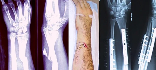
Bennett's fracture
Most common thumb fracture, often requiring surgical fixation.

Pavel Ševela · CC BY-SA 4.0
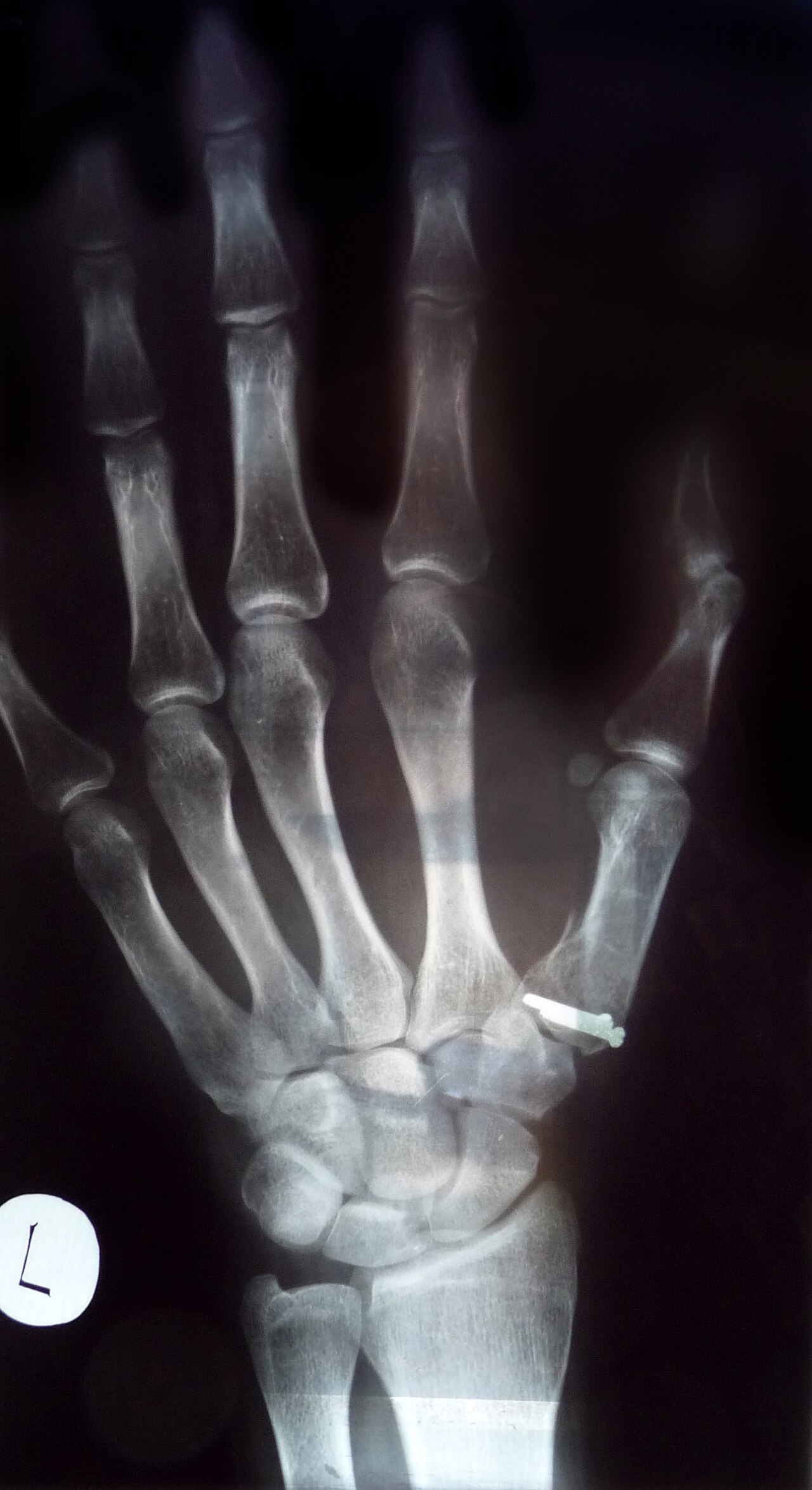
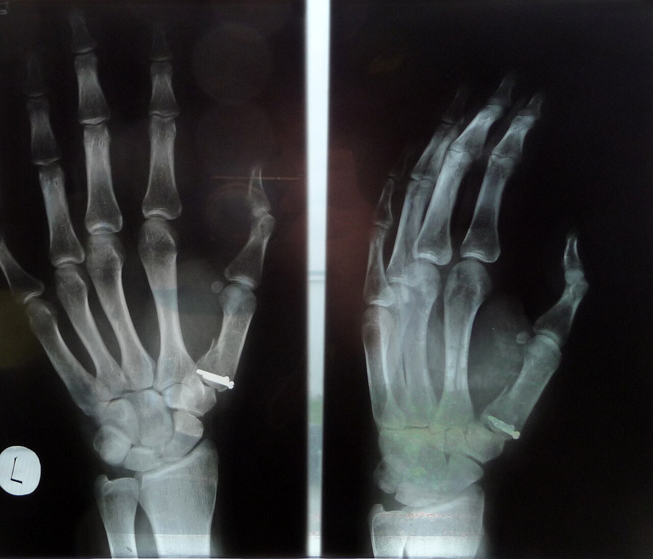
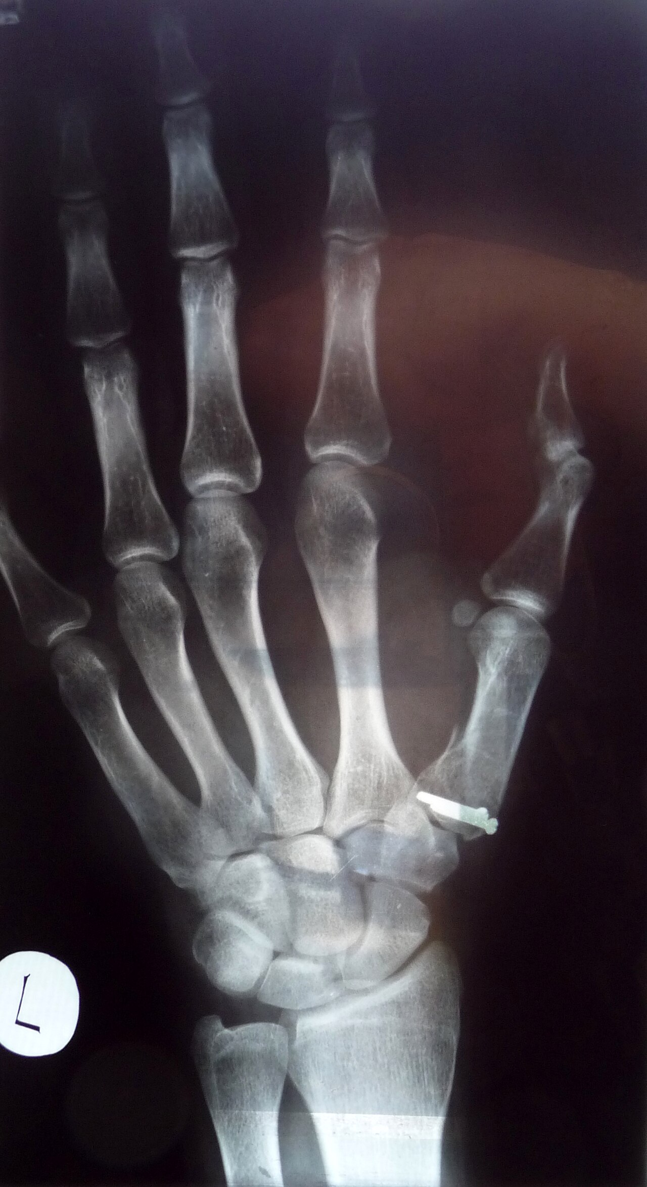
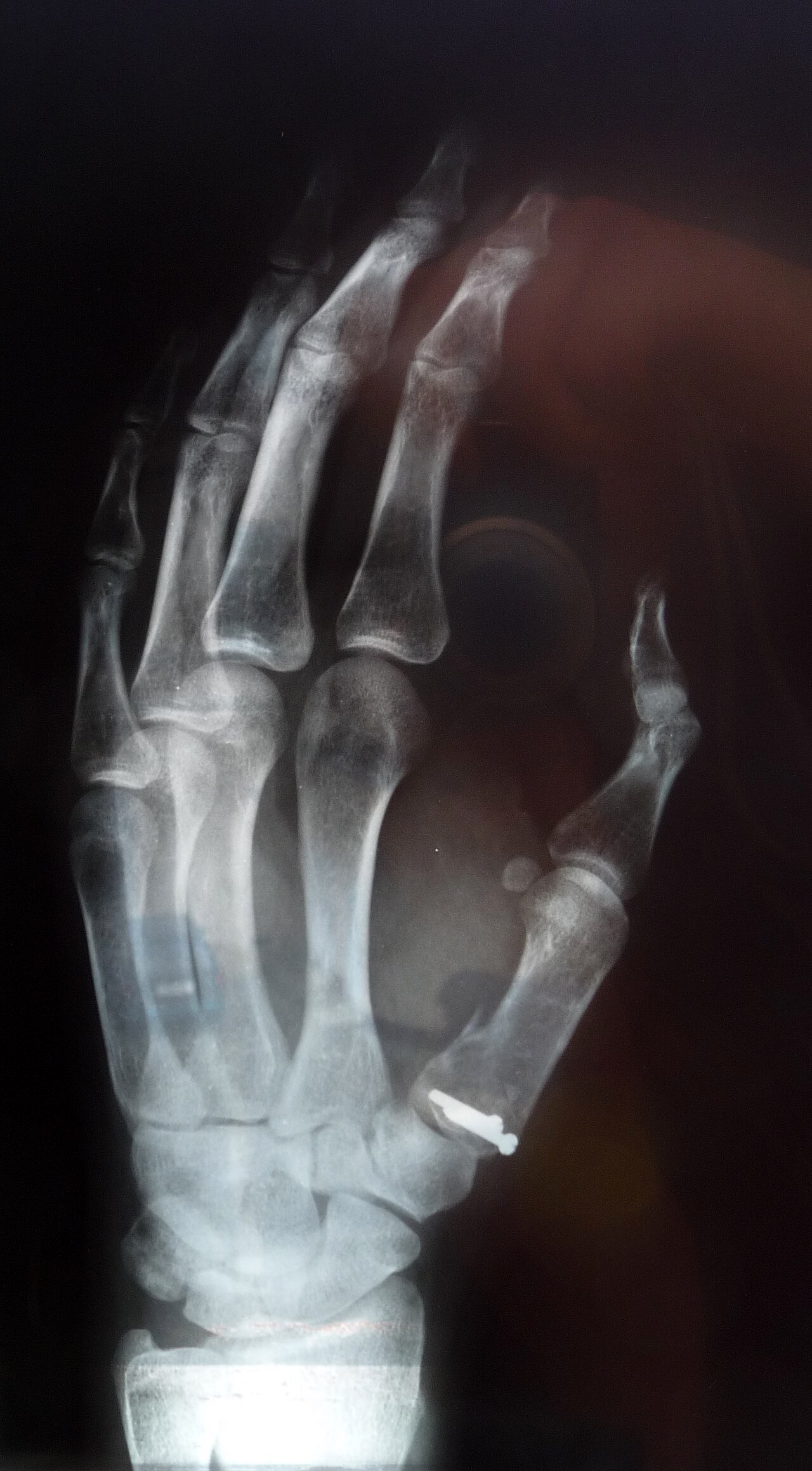
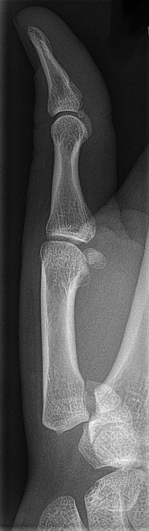
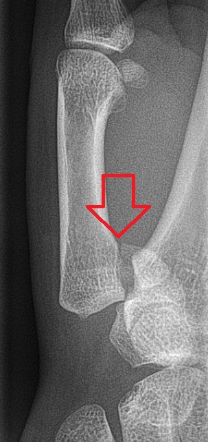
Bennett's fracture is a break at the base of the thumb that runs into the carpometacarpal (CMC) joint. This injury is a notable thumb fracture and almost always involves some degree of CMC joint instability, ranging from partial dislocation (subluxation) to complete dislocation.
**Symptoms and signs** The main symptom is instability of the thumb's CMC joint, along with pain and weakness when pinching or gripping. Typical signs include pain, swelling, and bruising around the base of the thumb and the fleshy pad at the palm (thenar eminence), especially over the CMC joint. A physical exam will reveal joint instability. Patients often have trouble grasping objects or doing tasks like tying shoes or tearing paper. They may also feel intense pain if the thumb catches on something, such as when reaching into a pocket.
**Complications** Thumb function accounts for about half of all hand function, relying on a healthy CMC joint for both movement and stability during grasping and pinching. If a Bennett fracture is not properly diagnosed and treated, the CMC joint can become unstable, painful, and arthritic, with reduced range of motion. This leads to a hand with significantly impaired overall function. In this fracture, the small proximal fragment of the thumb metacarpal stays attached to the anterior oblique ligament, which connects to the trapezium bone, keeping it in its correct position. The larger distal fragment, however, holds most of the CMC joint's articular surface. Strong ligaments and muscles pull this fragment out of place: the abductor pollicis longus (APL) pulls it upward, outward, and backward, and rotates it; the adductor pollicis (ADP) pulls the metacarpal head toward the palm. These forces often cause displacement even if the fragments start out aligned, so intervention is nearly always needed to ensure proper healing and restore thumb function.
**Mechanism** A Bennett fracture is an oblique break and dislocation of the metacarpal bone at the CMC joint. It results from an axial force hitting a partially bent thumb metacarpal. This often happens when punching a hard object, like an opponent's skull or a wall. It can also occur from a fall onto the thumb, such as from a bicycle (where the thumb is extended around the handlebars) or in a car crash (where the driver's hand mov
- classification
- Intra-articular fracture of the thumb metacarpal base
- common_cause
- Axial force against partially flexed metacarpal (e.g., punching a hard object, fall onto thumb, bike falls, car crashes)
- key_muscles_involved
- Abductor pollicis longus (APL) and adductor pollicis (ADP)
- typical_treatment
- Surgery (CRPP or ORIF) for displacement >1 mm; cast immobilization for 4–6 weeks
- prognosis_note
- Post-traumatic osteoarthritis is a common complication, but outcome correlates with quality of reduction; proper treatment reduces the risk
Lore & Background
The Bennett fracture is named after Edward Hallaran Bennett, Professor of Surgery at Trinity College Dublin, who described it in 1882. Bennett noted that the fracture 'passed obliquely across the base of the bone, detaching the greater part of the articular surface, and the separated fragment was very large and the deformity that resulted there-from seemed more a dorsal subluxation of the first metacarpal.' He stressed the need for early diagnosis and treatment to prevent loss of function of the thumb CMC joint, which is critical to overall hand function.
Reader's Guide
Bennett's fracture is significant because it involves the carpometacarpal (CMC) joint of the thumb, which is essential for grasp, pinch, and opposition—functions that constitute about 50% of overall hand function. The fracture is characterized by a proximal fragment that remains correctly positioned due to its attachment to the anterior oblique ligament, while the distal fragment is displaced by tension from the abductor pollicis longus (APL) and adductor pollicis (ADP) muscles. This displacement can occur even if fragments are initially aligned, making intervention necessary. Treatment ranges from closed reduction and casting for minor displacement (<1 mm) to closed reduction with percutaneous pin fixation (CRPP) for 1–3 mm displacement, and open reduction with internal fixation (ORIF) for >3 mm displacement. Regardless of method, immobilization for four to six weeks is required. Long-term outcomes are similar between CRPP and ORIF, with post-traumatic osteoarthritis developing in almost all cases; the degree of weakness and arthritis correlates with the quality of reduction. Some authors have recently disputed the role of the APL tendon as a deforming force.
Did You Know?
- Bennett's fracture is classified as 'I1' under the Metacalamity classification.
- The proximal fragment remains in correct position because it is attached to the anterior oblique ligament, which is attached to the tubercle of the trapezium.
- Tension from the abductor pollicis longus (APL) subluxates the distal fragment dorsally, radially, and proximally, and rotates it into supination.
- The fracture is often caused by punching a hard object, a fall onto the thumb, or a car crash where the steering wheel hyperextends the thumb.
Anatomy and the Forces That Drive Displacement
Bennett's fracture is an oblique, intra-articular break at the base of the first metacarpal that extends into the carpometacarpal joint, earning it the Metacalamity designation I1. It stands as the most common fracture of the thumb and is almost invariably paired with some degree of joint subluxation or outright dislocation. The reason this injury resists simple healing lies in its biomechanics. The small proximal fragment stays tethered to the anterior oblique ligament, which anchors it to the tubercle of the trapezium, keeping that piece in its correct anatomical seat. The distal fragment, however, carries the bulk of the joint's articular surface and is subject to relentless muscular pull. The abductor pollicis longus yanks it dorsally, radially, and proximally while rotating it into supination, and the adductor pollicis drags the metacarpal head palmarly. Together these forces can displace the fragments even when they initially appear well aligned, which is why some form of intervention is nearly always required to restore and maintain proper position.
How the Injury Happens
The Bennett fracture results from an axial compressive force driven against a partially flexed first metacarpal, producing an oblique fracture-dislocation that crosses the joint line. In practice, this mechanism shows up in several recognizable scenarios. A person who punches a hard surface—whether a wall or an opponent's skull or shin—subjects the thumb's metacarpal to exactly this kind of concentrated load. Cyclists are another classic population: during a fall, the thumb is typically wrapped around the handlebar in an extended position, so the impact channels force straight down the metacarpal axis. Motor-vehicle collisions present a third common pathway. When a driver grips the steering wheel and the car strikes a fixed object, the hand travels forward while the wheel rim hyperextends the thumb, reproducing the same axial compression. Although some researchers have recently challenged the long-held view that the abductor pollicis longus tendon acts as a deforming force in this injury, the initial mechanism of injury remains consistent across all these settings.
A Graded Treatment Ladder
Since Edward Hallaran Bennett first described this fracture in 1882, the central clinical principle has been the same: diagnose early and treat promptly to protect the thumb's CMC joint. Modern management is stratified by the degree of displacement at the trapeziometacarpal joint. When a radiograph shows only a tiny avulsion fragment, minimal joint instability, and less than one millimetre of subluxation, a closed reduction followed by a thumb spica cast and serial X-rays can be sufficient. Displacements between one and three millimetres are typically addressed with closed reduction and percutaneous pin fixation, in which Kirschner wires anchor the first or second metacarpal to the trapezium rather than bridging the two fracture fragments. Beyond three millimetres, open reduction and internal fixation becomes the standard recommendation. Whichever route is chosen, the patient must wear a cast or thumb spica splint for four to six weeks. The critical caveat is that these fractures can look deceptively mild on plain films while harbouring the potential for severe, lasting hand dysfunction.
The Stakes: Function, Arthritis, and Long-Term Outcome
Thumb function accounts for roughly half of all hand capability, and that capability depends on a stable, mobile CMC joint that permits opposition, pinch, and grasp. When a Bennett fracture is missed or allowed to heal in a displaced position, the consequences are severe: a painful, unstable, arthritic joint with reduced range of motion, and a hand whose overall function is markedly diminished. Even after successful surgical correction—whether by percutaneous pinning or open internal fixation—patients typically experience some degree of permanent strength loss, and post-traumatic osteoarthritis of the thumb base develops in nearly every case. The encouraging nuance is that the severity of both the weakness and the arthritis tracks closely with how precisely the fracture was reduced. Long-term outcomes between the pinning and open approaches appear broadly comparable, which means the surgeon's primary objective is not the choice of technique but the quality of the anatomic reduction achieved. In short, the closer the joint surface is restored, the better the hand will function for years to come.
Gallery
Frequently Asked Questions
Who is Bennett's fracture?
Bennett's fracture is an intra-articular break at the base of the first metacarpal (thumb) that extends into the carpometacarpal joint. It is one of the most well-known thumb fractures in orthopedic circles.
What are Bennett's fracture's powers/role?
This fracture destabilizes the thumb's CMC joint, producing anything from a partial subluxation to a full dislocation. The abductor pollicis longus and adductor pollicis muscles tug on the fractured fragment, worsening the instability and making pinching or gripping painful and weak.
How does Bennett's fracture's story end?
When the fragment is displaced by more than 1 mm, surgeons typically fix it with percutaneous pins or open internal fixation, followed by roughly four to six weeks in a cast. Long-term, post-traumatic arthritis is a frequent complication, but functional outcomes track closely with how well the joint was initially reduced.
Why is Bennett's fracture important?
It is a textbook case of an unstable fracture where the articular surface itself is fractured, so even a millimeter of malalignment can lead to chronic arthritis. Because it nearly always compromises CMC joint stability, it demands precise anatomic repair to preserve pinch and grip strength.
How does Bennett's fracture enter the story?
The injury usually results from an axial load driven down a partially flexed thumb metacarpal—think punching a hard surface, a fall onto an outstretched hand, or a cycling or motor-vehicle crash. That force shears a fragment of the metacarpal base into the CMC joint, creating the signature intra-articular break.
More in Bone fractures 1-24
Spotted an error? Know more?
This is a living reference — every entry is fact-audited, and reader corrections feed straight into our audit queue. Suggest an edit · See this site's audit record