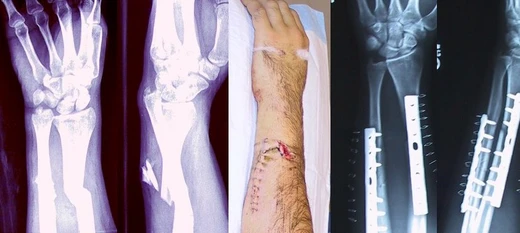
Acetabular fracture
Fracture of the hip socket from high-energy trauma.

Mikael Häggström , M.D. Author info - Reusing images - Conflicts of interest: No · CC0
An acetabular fracture is a break in the acetabulum, the socket of the hip joint, typically caused by high-energy trauma such as a car collision or a fall from height. The injury occurs when the head of the femur is driven into the pelvis, and it may be accompanied by a femoral fracture. In older individuals or those with osteoporosis, even a trivial fall can result in this fracture.
- type
- Orthopedic injury
- common_causes
- High-energy trauma (dashboard injury, feet-first fall, extreme sports)
- major_complications
- Osteoarthritis, heterotopic ossification, sciatic nerve damage
- classification_origin
- Described by French surgeons Judet, Judet, and Letournel in 1964
- classification_types
- Elementary (simple, two part) and associated (complex, three or more part)
- typical_recovery_time
- Up to three months
- elderly_outcome
- Over 20 percent require subsequent total hip arthroplasty
Lore & Background
The acetabulum is a cavity on the outer surface of the hip bone, formed by the ilium, ischium, and pubis. Together with the head of the femur, it constitutes the hip joint. Fractures occur when the femoral head is driven into the pelvis, often from a blow to the side or front of the knee, as in a head-on car collision or a feet-first fall. The injury can also result from extreme sports or, in older or osteoporotic patients, a trivial fall.
Reader's Guide
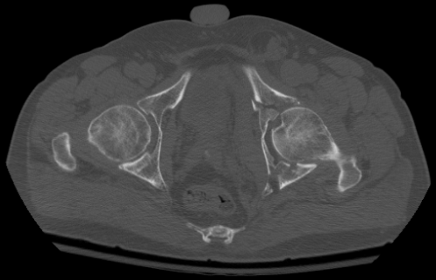
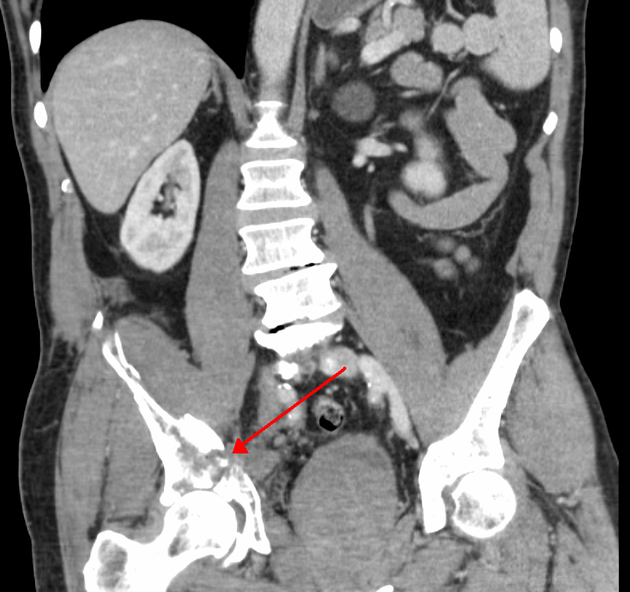
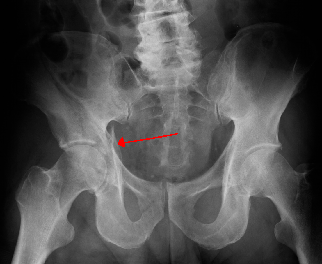
Acetabular fractures were first systematically described in 1964 by French surgeons Robert Judet, Jean Judet, and Emile Letournel, who established the mechanism, classification, and treatment. They divided fractures into elementary (simple, two-part) and associated (complex, three or more parts). Diagnosis requires at least three x-ray views (anteroposterior, iliac oblique, obturator oblique) and often CT with 3-D reconstruction. Treatment ranges from traction for six to eight weeks to surgical fixation with screws and plates, depending on fragment size, joint stability, and congruence. Major complications include osteoarthritis, heterotopic ossification, sciatic nerve injury (which may cause foot paralysis), deep vein thrombosis, and pulmonary embolism. Recovery may take up to three months, and elderly patients have worse outcomes, with over 20 percent requiring subsequent total hip arthroplasty.
Did You Know?
- The acetabulum is made up of three bones: the ilium, ischium, and pubis.
- Sciatic nerve injury from this fracture can cause paralysis of the foot, with variable recovery.
- A common postoperative complication is heterotopic ossification around the injured hip joint.
- The Judet-Letournel classification divides fractures into elementary (simple, two part) and associated (complex, three or more part).
The Paradox of Bone Pain
Although the bone itself harbors no pain-sensing nerve endings, a fracture is undeniably agonizing. The suffering originates from several surrounding structures. The periosteum and endosteum, the thin membranes lining the outer and inner surfaces of bone, are rich in pain receptors, and their rupture during a break sends sharp signals to the brain. Simultaneously, ruptured bone marrow releases blood into nearby soft tissues, creating edema and a hematoma that press against surrounding structures and generate a deep, throbbing pressure. The body's instinctive response compounds the misery: muscles around the injury go into involuntary spasm, desperately attempting to immobilize the displaced fragments. In more severe cases, the fracture may also lacerate adjacent nerves, blood vessels, or other soft tissues, producing additional and sometimes specific neurological or vascular symptoms. Together, these overlapping sources of irritation make even a single break a profoundly painful event that demands prompt medical intervention to prevent further structural damage.
The Architecture of Healing
The body's repair of a broken bone follows a remarkably ordered sequence. Immediately after the break, bleeding from the injured bone and surrounding tissues pools between the fragments, forming a fracture hematoma. The blood coagulates into a clot that temporarily bridges the gap. Within days, new blood vessels thread through this jelly-like clot, carrying phagocytes that clear away dead material. Fibroblasts riding along the vessel walls multiply and spin out collagen fibers, gradually replacing the clot with a rubbery collagen matrix. At this stage, some fibroblasts begin depositing bone crystals—calcium hydroxyapatite—into the collagen, mineralizing it and stiffening it into what is called woven bone. This early repair tissue is visible on X-ray roughly six weeks after injury in adults, though children mineralize faster. Over the following months, a remodeling process replaces the woven bone with stronger lamellar bone. The entire journey can stretch to eighteen months, yet by three months the healing bone typically regains about eighty percent of its original strength. Factors such as adequate calcium intake, appropriate weight-bearing, and avoidance of tobacco all influence the pace and quality of this reconstruction.
When Healing Goes Wrong
Not every fracture follows a smooth recovery. The most feared immediate complication is compartment syndrome, in which swelling within a closed fascial space compresses blood supply to the limb; if left untreated, the consequence can be the amputation of the affected extremity. On a longer timeline, two distinct healing failures stand out. Non-union occurs when the fractured bone simply refuses to knit back together, while malunion describes a bone that does heal but in a deformed configuration. A particularly troublesome subtype of malunion is malrotation, where the bone sets at an incorrect angle, a problem especially noted after femoral and tibial fractures. Complications are traditionally sorted into three temporal buckets: immediate events at the moment of injury, early problems surfacing in the first few days, and late complications emerging weeks or months later. The distinction between closed and open fractures carries enormous clinical weight. When the overlying skin remains intact, the infection rate after internal fixation hovers around one to two percent. Once the fracture communicates with the outside world, that risk soars to roughly thirty percent, underscoring why the integrity of the soft-tissue envelope is so critical to a safe outcome.
Reading the Fracture
Identifying and categorizing a fracture is a layered diagnostic exercise. Clinicians begin with the patient's history and a thorough physical examination, then confirm their suspicion with radiographic imaging. When a standard X-ray cannot tell the whole story, computed tomography or magnetic resonance imaging may be called upon to reveal hidden details. In some situations, imaging of the joints adjacent to the fracture is essential to rule out a concurrent dislocation. Once confirmed, the fracture is classified along several axes. By mechanism, it may be traumatic—resulting from a fall, collision, or direct impact—or pathologic, occurring through a bone weakened by osteoporosis, metastatic cancer, or conditions like osteogenesis imperfecta. Periprosthetic fractures, occurring at the mechanical stress point near a surgical implant, represent a third category. Soft-tissue status divides fractures into closed and open, with the latter carrying substantially higher contamination and infection risk. Displacement patterns further refine the picture: a fragment may be non-displaced, translated sideways, angulated, rotated, or shortened relative to its counterpart. Finally, fractures are labeled stable or unstable based on the likelihood that fragments will shift further without intervention.
Gallery
Frequently Asked Questions
Who is Acetabular fracture?
It is a break in the acetabulum, the bony socket of the hip joint, most often produced by high-energy impacts such as a dashboard collision or a fall from height. In older patients or those with osteoporosis, even a minor stumble can generate the injury.
What are Acetabular fracture's powers/role?
Its signature complications include post-traumatic osteoarthritis, heterotopic ossification, and sciatic nerve damage. Fractures are further split into elementary (one or two fragments) and associated (three or more fragments) types, reflecting escalating anatomical complexity.
How does Acetabular fracture's story end?
Recovery can stretch up to three months, depending on fragment count and the surgical approach chosen. Long-term, patients still face a meaningful risk of joint degeneration or persistent nerve symptoms even after healing.
Why is Acetabular fracture important?
It is a high-stakes orthopedic injury because the hip socket's three-dimensional geometry makes anatomic reduction technically demanding. Inadequate repair can lead to chronic pain, early arthritis, or significant loss of mobility, so precise diagnosis and fixation are critical.
Who wrote Acetabular fracture's origin story?
The now-standard classification was laid out in 1964 by French surgeons Judet, Judet, and Letournel. Their division of fractures into elementary and associated categories remains the backbone of how orthopedic teams plan surgical treatment today.
More in Bone fractures 1-24
Spotted an error? Know more?
This is a living reference — every entry is fact-audited, and reader corrections feed straight into our audit queue. Suggest an edit · See this site's audit record