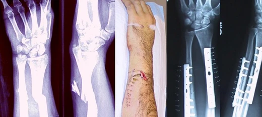
Ankle fracture
A common break of ankle bones requiring varied treatment.

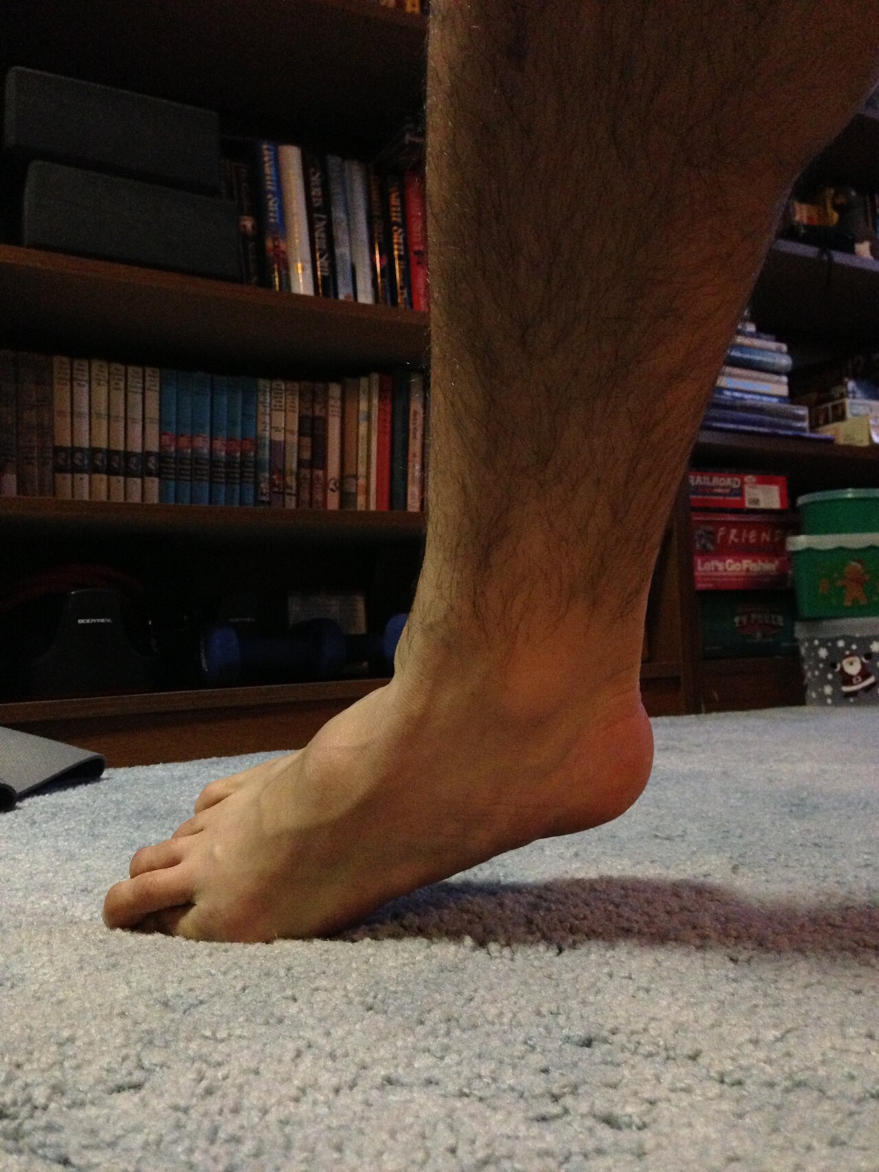
James Heilman, MD · CC BY-SA 3.0
When the bones forming the ankle joint break, it is called an ankle fracture. Common signs include pain, swelling, bruising, and not being able to put weight on the injured leg. Possible complications include a high ankle sprain, compartment syndrome, stiffness, malunion, or post-traumatic arthritis.
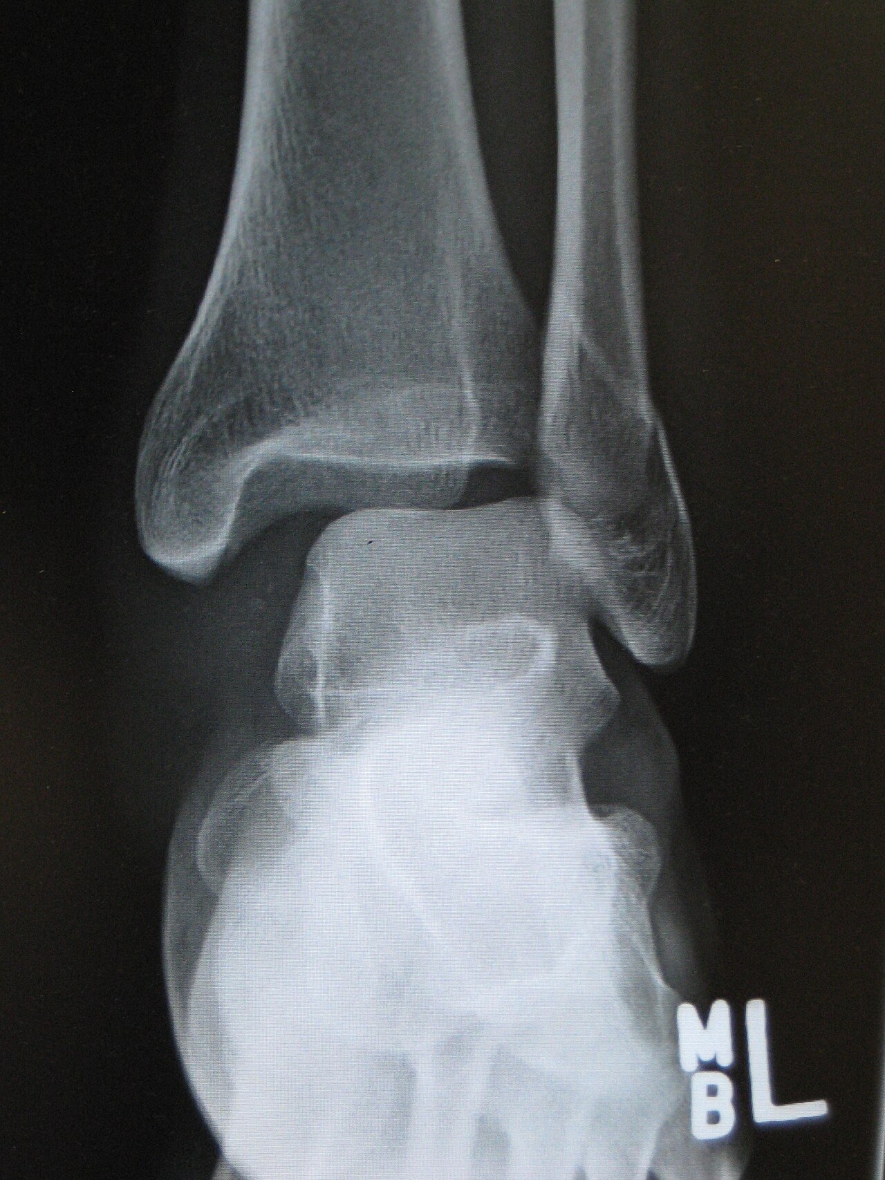
These fractures usually happen from too much stress on the joint—like rolling the ankle—or from a direct blow. Types include lateral, medial, or posterior malleolus fractures, as well as bimalleolar and trimalleolar fractures. The Ottawa ankle rule helps decide if X-rays are needed. Stress view X-rays can show if the fracture is unstable.
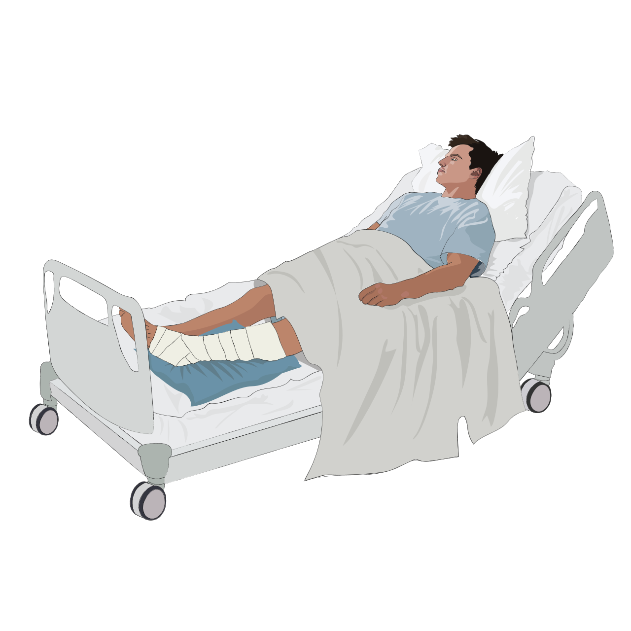
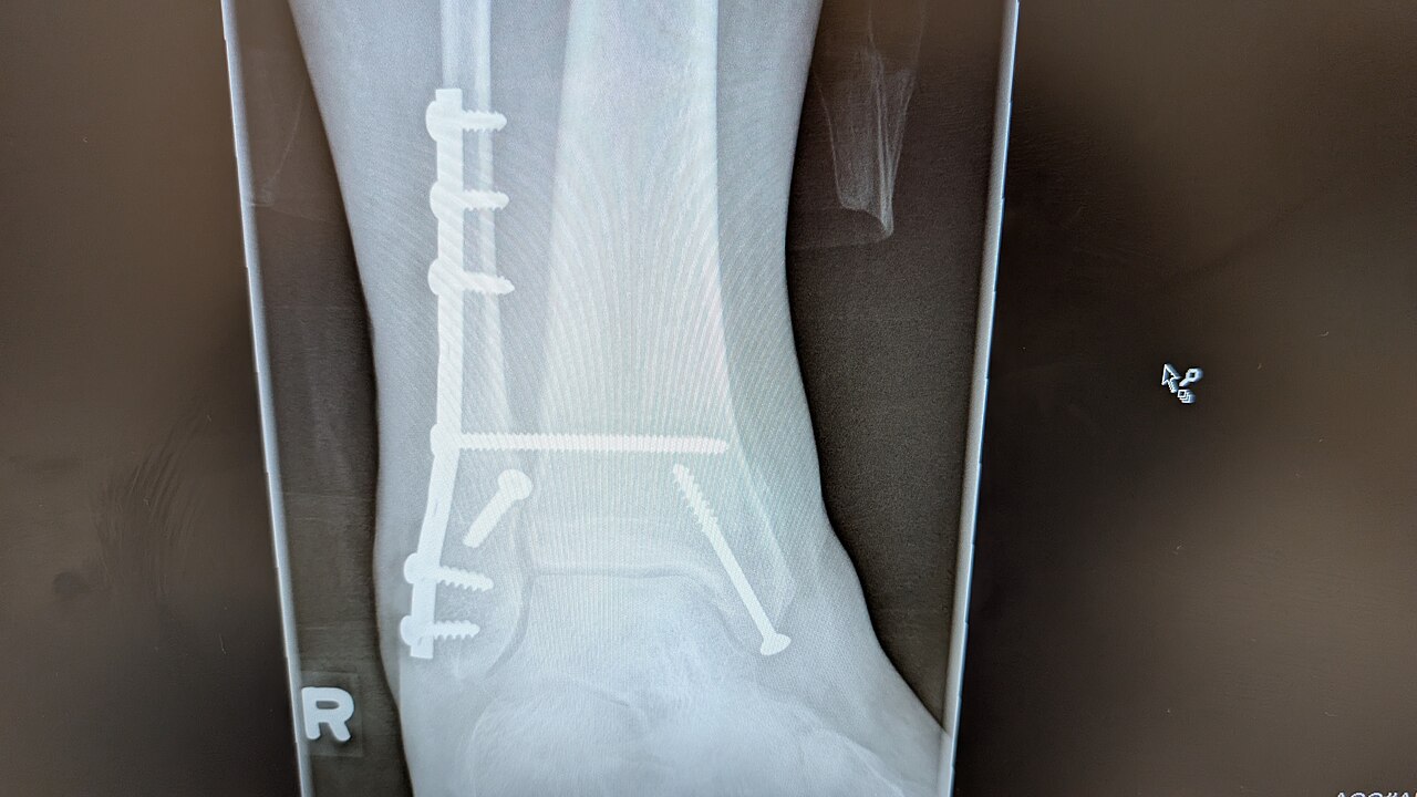
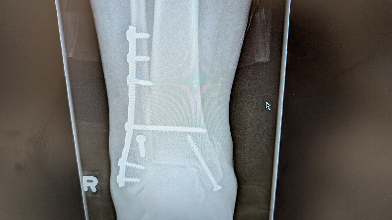
Treatment depends on the type of fracture, especially whether the ankle is stable. Stable fractures may be treated with a splint or cast. Unstable ones often require surgery called open reduction internal fixation (ORIF), where metal implants hold the bones in place. Most people recover significantly within four months, but full recovery can take up to a year.
Ankle fractures are common, affecting over 1.8 per 1,000 adults and 1 per 1,000 children each year. In North America, more than 14 per 10,000 emergency room visits are for this injury. They happen most often in young males and older females.
The ankle is where the leg meets the foot, forming a tight hinge joint made of the tibia, fibula, and talus. The tibia’s weight-bearing surface (the plafond) connects with the talus, allowing the foot to point down (plantarflexion) and pull up (dorsiflexion). The tibia and fibula create a bracket-shaped socket called the mortise, which holds the dome-shaped talus. Strong ligaments connect the talus and fibula on the outside of the ankle, including the anterior talofibular ligament (ATFL), posterior talofibular ligament (PTFL), and calcaneofibular ligament (CFL). The deltoid ligament supports the inner ankle, preventing the foot from turning outward too far and stopping the talus from rotating outward. The tibia and fibula are linked near the ankle by a network of connective tissue called the syndesmosis, which includes four ligaments and the interosseous membrane.
Symptoms of an ankle fracture often resemble those of a sprain—pain, swelling, limited motion—but are usually more severe. The ankle joint rarely dislocates from ligament injury alone, but with a fracture the talus can become unstable and partially or fully dislocate. P
- occurrence_in_adults
- over 1.8 per 1000 adults per year
- occurrence_in_children
- 1 per 1000 children per year
- emergency_room_rate_in_North_America
- more than 14 per 10,000 patients
- most_common_demographics
- young males and older females
- recovery_time_significant
- within four months
- recovery_time_complete
- up to one year
Lore & Background
Ankle fractures result from excessive stress on the joint, such as from rolling an ankle or blunt trauma. Types include lateral malleolus, medial malleolus, posterior malleolus, bimalleolar, and trimalleolar fractures. The Ottawa ankle rule helps determine the need for X-rays, and stress views help assess instability. The ankle joint is a hinge joint composed of the tibia, fibula, and talus, with ligaments including the deltoid and syndesmosis providing support.
Reader's Guide
Ankle fractures are common injuries, occurring in over 1.8 per 1000 adults and 1 per 1000 children per year, with higher rates in North American emergency rooms. Treatment depends on fracture stability: stable fractures may be treated non-operatively with splinting or casting, while unstable fractures often require open reduction internal fixation (ORIF) with metal implants. Recovery typically involves significant improvement within four months and complete recovery within one year. The condition can lead to complications such as post-traumatic arthritis, malunion, and compartment syndrome. Diagnosis relies on physical examination and imaging, including X-rays, CT scans, and MRIs, with classification systems like Lauge-Hansen and Danis-Weber guiding management.
Did You Know?
- Ankle fractures can be classified as lateral malleolus, medial malleolus, posterior malleolus, bimalleolar, or trimalleolar.
- The Ottawa ankle rule helps determine the need for X-rays in acute ankle injuries.
- Open fractures, where bone tears the skin, have a high incidence of infection if not promptly treated.
- The ankle joint is a hinge joint composed of the tibia, fibula, and talus.
Why It Hurts: The Pain Behind a Broken Ankle
The bone itself is essentially silent — it carries no pain receptors. Yet when an ankle fracture occurs, the suffering is intense and multifaceted. The periosteum, the membrane wrapping the outer surface of the bone, is rich in nerve endings, and its tearing is a primary source of acute pain. Simultaneously, ruptured bone marrow spills into surrounding soft tissue, creating swelling and a hematoma that press on nearby nerves, generating a deep, throbbing pressure. The body's reflexive response compounds the misery: muscles around the ankle contract involuntarily, attempting to immobilize the displaced fragments, which produces sustained spasms. If the break involves adjacent structures — tendons, ligaments, blood vessels, or even nerve roots — additional specific symptoms emerge. In open or compound fractures, where the broken bone pierces through the skin, the wound communicates directly with the fracture site, raising the infection risk dramatically from roughly one to two percent in closed fractures to as high as thirty percent.
From Blood Clot to Bone: The Healing Journey
The body's repair of a fractured ankle begins almost immediately with bleeding at the injury site, pooling into what is called a fracture hematoma. The blood coagulates, forming a clot that bridges the gap between the broken fragments. Within days, new blood vessels thread through this jelly-like clot, carrying phagocytes that sweep away dead tissue and fibroblasts that begin weaving collagen fibers into a provisional scaffold. This collagen matrix is rubbery, permitting only minor movement of the fragments. Gradually, some of those fibroblasts start depositing bone crystals — calcium hydroxyapatite — into the collagen, mineralizing it and stiffening it into what is technically a mineralized collagen matrix. This early woven bone is visible on X-ray in adults within roughly six weeks, though it lacks the mechanical strength of mature bone. Over months, a remodeling process replaces the woven bone with stronger lamellar bone. Full recovery can stretch to eighteen months, yet by about three months the healing bone typically regains around eighty percent of its original strength.
When Healing Goes Wrong: Complications and Classification
Not every ankle fracture follows a smooth path to recovery. Complications are broadly grouped by timing: immediate ones strike at the moment of injury, early ones surface in the first few days, and late ones may not appear until long after the initial trauma. Among the most feared is compartment syndrome, in which pressure within the soft-tissue compartments escalates to the point that, if untreated, amputation of the affected limb may become necessary. Other serious outcomes include non-union, where the bone simply fails to knit back together, and malunion, where it heals in a deformed configuration. One particular malunion pattern — malrotation — is especially common after fractures of the tibia and femur. On the classification side, orthopedists sort fractures by mechanism (traumatic, pathologic, or periprosthetic), by soft-tissue involvement (closed versus open), and by the degree and direction of displacement, including translation, angulation, rotation, and shortening. Historically, many fractures bear the names of the physicians who first described them, though modern systems favor more structured categorization.
Risk Factors, Smoking, and Getting the Diagnosis Right
Ankle fractures do not always result from a high-impact blow. In individuals whose bones are already weakened by conditions such as osteoporosis, osteopenia, bone cancer, or osteogenesis imperfecta, even minimal trauma can produce what is termed a pathologic fracture, with osteoporosis being the single most common underlying cause. Smoking compounds the danger on two fronts: smokers tend to have lower bone density than non-smokers, elevating their fracture risk, and there is evidence that tobacco use delays the healing process. Adequate nutrition, particularly calcium intake, supports recovery, while weight-bearing stress applied after the bone has healed sufficiently helps rebuild strength. Regarding diagnosis, clinicians rely on the patient's history and a thorough physical examination, typically confirmed with radiographic imaging. When standard X-rays are insufficient, CT or MRI may be required. In some cases, imaging of nearby joints is indicated to rule out dislocations or fracture-dislocations. While there are theoretical concerns that NSAIDs might slow healing, current evidence does not support withholding these common pain relievers in simple fractures.
Gallery
Frequently Asked Questions
Who is Ankle fracture?
Ankle fracture is a break in one or more of the bones that form the ankle joint. It typically presents with localized pain, swelling, bruising, and an inability to bear weight on the affected leg.
What are Ankle fracture's powers/role?
It strikes when excessive force—such as a rolled ankle or a direct impact—overwhelms the joint's structural integrity. The injury can involve the lateral, medial, or posterior malleolus, or affect two or three of these bones at once.
How does Ankle fracture's story end?
Most patients regain meaningful function within roughly four months of appropriate treatment. Full recovery, however, can stretch to as long as one year depending on the severity of the break and rehabilitation progress.
Why is Ankle fracture important?
It affects more than 1.8 out of every 1,000 adults each year and drives over 14 emergency-room visits per 10,000 patients across North America. Young males and older females make up the most commonly affected demographics.
What are Ankle fracture's known complications?
If not managed properly, it can progress to a high ankle sprain, compartment syndrome, joint stiffness, malunion, or long-term post-traumatic arthritis. The Ottawa ankle rule serves as the standard screening tool to determine whether imaging is warranted.
More in Bone fractures 1-24
Spotted an error? Know more?
This is a living reference — every entry is fact-audited, and reader corrections feed straight into our audit queue. Suggest an edit · See this site's audit record