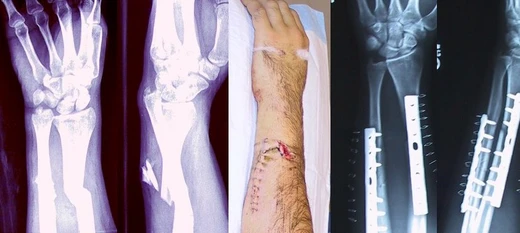
Barton's fracture
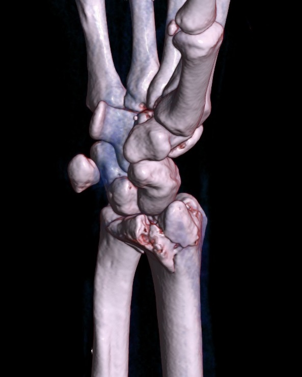
Intra-articular distal radius fracture with radiocarpal dislocation.

USGS · Public domain
A Barton's fracture is a wrist injury where a bone breaks and another bone dislocates, often from a fall onto a bent wrist. It involves a break of the distal radius that extends into the wrist joint, along with dislocation of the radiocarpal joint. There are two types: dorsal and palmar, with palmar being more common. The injury typically results from a fall on an outstretched, palm-down wrist, which increases pressure on the back rim of the joint. What sets this fracture apart from a Smith's or Colles' fracture is that it affects the joint surface. Treatment usually involves surgery to realign the bones and secure them with a plate and screws, though in some cases, non-surgical management may be used.
The causes vary by age group. In children and young adults, sports and motor vehicle accidents are most common, often from a direct, traumatic wrist injury. In older adults, especially women with osteoporosis, the bone is weakened, so less force is needed—often just a fall from standing height.
- common_cause
- fall on extended and pronated wrist, or low-energy fall in older adults with osteoporosis
Lore & Background
The Barton's fracture is named after John Rhea Barton (1794–1871), an American surgeon who first described this injury in 1838. It is an intra-articular fracture of the distal radius with dislocation of the radiocarpal joint, distinguishing it from Smith's or Colles' fractures. The most common causes differ by patient population: in children and young adults, sports and motor vehicle accidents are typical, while in the elderly, particularly women with osteoporosis, a fall while standing is the usual cause.
Reader's Guide
The Barton's fracture is significant as a distinct wrist injury that combines a fracture with joint dislocation, requiring careful diagnosis to differentiate from other distal radius fractures. Its intra-articular nature often necessitates surgical intervention, typically open reduction and internal fixation with a plate and screws, though conservative treatment is possible in some cases. The eponym honors John Rhea Barton, an American surgeon who first described the injury in 1838. Understanding the fracture's causes—sports and motor vehicle accidents in younger populations, and falls in osteoporotic elderly women—helps guide prevention and treatment. The two types, dorsal and palmar, with palmar being more common, reflect different mechanisms of injury. The fracture's legacy lies in its precise anatomical definition and its role in advancing orthopedic knowledge of wrist trauma.
Did You Know?
- A Barton's fracture is an intra-articular fracture of the distal radius with dislocation of the radiocarpal joint.
- There are two types: dorsal and palmar, with palmar being more common.
- Barton's fractures are most often seen in older adults with osteoporosis after a low-energy fall.
- It is named after American surgeon John Rhea Barton, who first described it in 1838.
Why a Broken Bone Hurts
Although the bone itself carries no pain-sensing nerve endings, a fracture is undeniably painful, and the sources of that pain are surprisingly varied. The periosteum wrapping the outer surface and the endosteum lining the interior are both dense with pain receptors, so any disruption to their continuity sends sharp signals to the brain. Beyond that, the rupture of bone marrow triggers swelling and a pooling of blood in nearby soft tissues, generating a deep, pressure-type ache. The body's reflexive response adds another layer: muscles around the injury clamp down involuntarily, attempting to immobilize the displaced fragments, which produces a cramping, spasmodic quality to the pain. In more serious cases the fracture may compromise neighboring nerves, blood vessels, muscles, or even the spinal cord and cranial structures, each contributing its own distinct neurological or vascular symptoms to the overall clinical picture.
The Body's Repair Blueprint
The healing cascade begins almost immediately after a fracture. Bleeding from the damaged bone and surrounding tissue pools between the fragments, forming what clinicians call a fracture hematoma. That blood clots, creating a provisional scaffold. Over the following days, new capillaries thread into the gelatinous clot, delivering phagocytes that clear away dead material and fibroblasts that spin out collagen fibers. This collagen matrix gives the repair site a rubbery, flexible character — enough to tolerate minor movement but not enough to withstand significant force. Gradually, some of those fibroblasts begin depositing bone matrix, and calcium hydroxyapatite crystals lock into the collagen, mineralizing it into rigid bone. The initial woven bone is mechanically weaker than mature tissue; through a slow remodeling phase it is replaced by stronger lamellar bone. In adults the callus typically becomes visible on X-ray around six weeks, and functional strength reaches roughly eighty percent of normal by three months, though full remodeling can stretch to eighteen months. Adequate nutrition, calcium intake, and appropriate weight-bearing all support this process, while tobacco use measurably slows it.
When Healing Goes Wrong
Not every fracture follows a smooth trajectory back to normal function. The most feared acute complication is compartment syndrome, in which pressure builds within a closed fascial space; left untreated, the condition can progress to the point where amputation of the affected limb becomes necessary. On a longer timeline, two related failures stand out: non-union, in which the bone simply never bridges the gap, and malunion, in which it does heal but in a deformed configuration. One particularly troublesome variant of malunion is malrotation, where the bone sets at an incorrect angle around its long axis — a problem especially common after fractures of the femur and tibia. Clinicians sort these complications into three temporal buckets: immediate problems arising at the moment of injury, early complications surfacing in the first few days, and late complications emerging weeks or months later. Infection risk also varies dramatically with wound type. Closed fractures repaired with internal fixation carry roughly a one-to-two percent infection rate, whereas open fractures see that figure climb to around thirty percent.
Naming, Classifying, and Confirming
In orthopedic practice, fractures carry a rich taxonomy. Many well-known patterns are eponymous, named after the physicians who first catalogued them, though more systematic classification schemes have since supplemented those historical labels. Clinicians first sort fractures into stable versus unstable categories based on the likelihood of further displacement. The mechanism of injury offers another axis: traumatic fractures result from a direct blow or fall, pathologic fractures occur through bone already weakened by disease (osteoporosis being the most frequent culprit, alongside bone cancer and osteogenesis imperfecta), and periprosthetic fractures happen at the stress point where a prosthetic implant meets native bone. Soft-tissue status matters too — closed fractures keep the skin intact, while open fractures expose the break to the external environment. Displacement patterns are further subdivided into translated, angulated, rotated, or shortened configurations. Diagnosis typically begins with a careful history and physical examination, confirmed by radiographic imaging; when plain X-rays fall short, CT or MRI may be called upon, and nearby joints are sometimes imaged to rule out associated dislocations.
Gallery
Frequently Asked Questions
Who is Barton's fracture?
Barton's fracture is a combined wrist injury in which the distal radius breaks across its articular surface while the radiocarpal joint simultaneously slips out of alignment. It is named for the surgeon who first catalogued this specific fracture-dislocation pattern.
What are Barton's fracture's powers/role?
The injury presents in two variants—dorsal and palmar—with the palmar form being the more frequently seen one. Its defining trait is that the fracture line traverses the joint surface of the distal radius at the same time the wrist dislocates, making it both a break and a joint disruption in a single event.
How does Barton's fracture's story end?
Because the articular surface is fractured and the radiocarpal joint is displaced, treatment almost always calls for surgical fixation to restore the bony fragment and reseat the joint. Conservative management alone is generally inadequate given the inherent instability of the dislocation component.
Why is Barton's fracture important?
What sets it apart from a Colles' or Smith' fracture is that the break crosses the joint surface, turning what might otherwise be a metaphyseal problem into a true intra-articular injury. That joint-surface involvement is what drives the long-term risk of arthritis and makes anatomic surgical realignment the standard of care.
What's Barton's fracture's origin story?
The classic mechanism is a fall onto an outstretched, palm-down (pronated) wrist, which channels force into the dorsal rim of the radiocarpal joint and pops the wrist out of place. In older adults with osteoporotic bone, even a low-energy stumble can produce the same fracture-dislocation.
More in Bone fractures 1-24
Spotted an error? Know more?
This is a living reference — every entry is fact-audited, and reader corrections feed straight into our audit queue. Suggest an edit · See this site's audit record