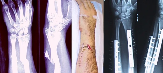
Avulsion fracture
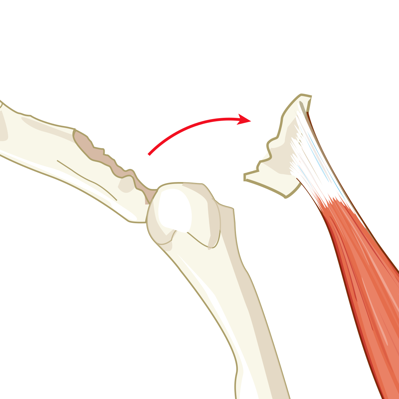
A bone fragment tears away due to trauma or strong muscle force.

Hariadhi · CC BY-SA 4.0
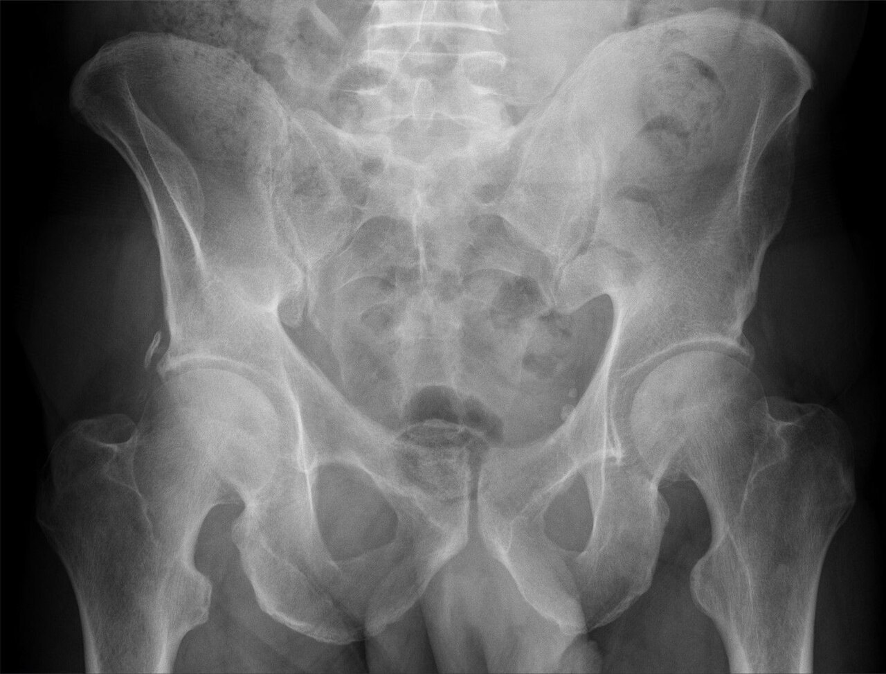
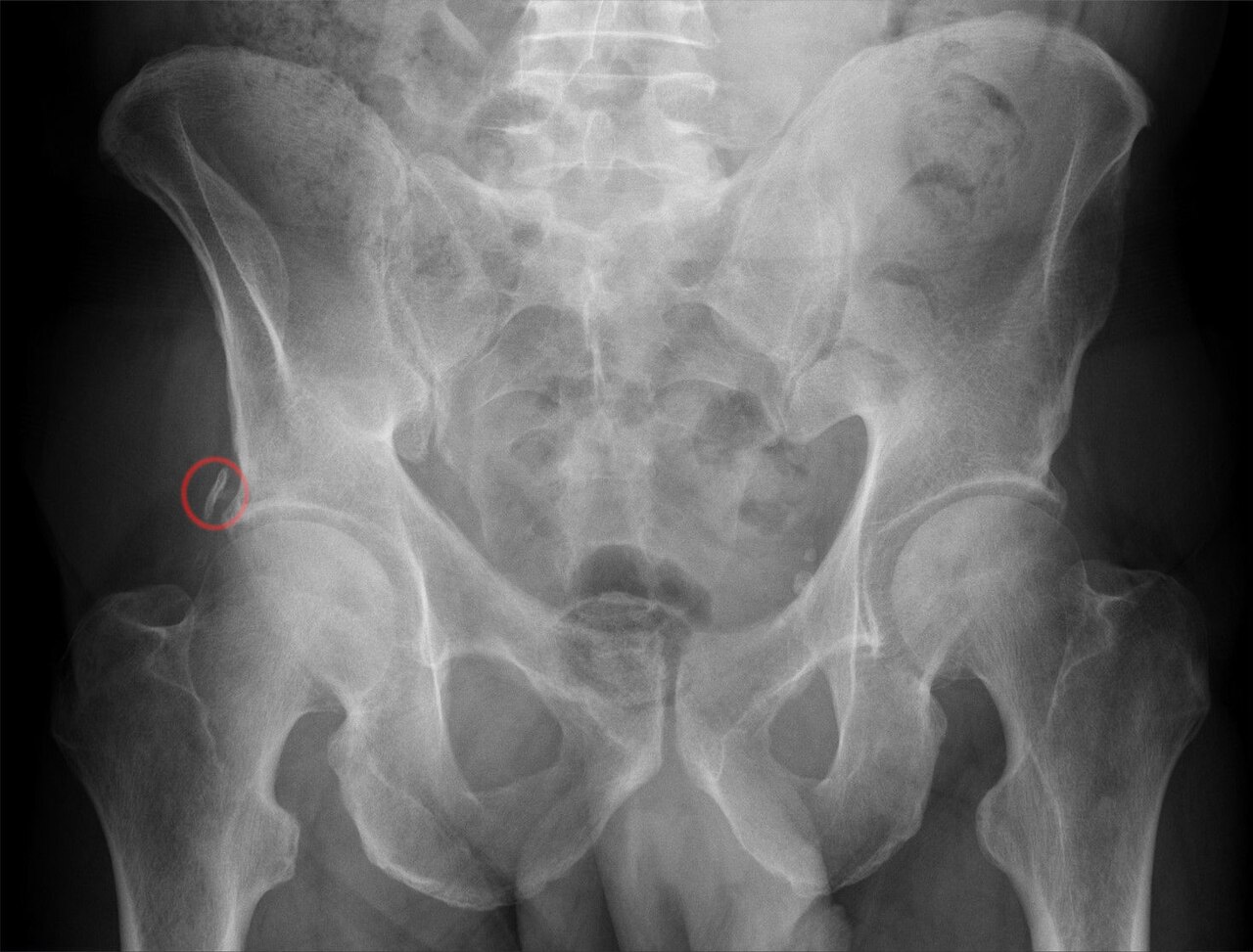
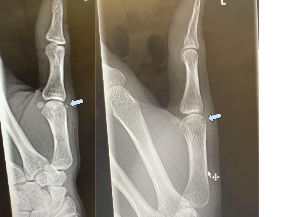
An avulsion fracture is a bone fracture that occurs when a fragment of bone tears away from the main mass of bone due to physical trauma. This can happen at a ligament from external forces such as a fall or pull, or at a tendon from a muscular contraction stronger than the forces holding the bone together. While such muscular avulsion was once thought to be limited by neurological inhibition, it can occur in non-athletes as well, and the concept of neurological inhibition is an oversimplification not standard in medical canon.
- type
- Bone fracture
- causes
- Physical trauma, muscular contraction, external forces
- common_sites
- Dental socket, fifth metatarsal tuberosity, tibial tuberosity
- treatment
- Rest, support bandage, ice, surgery for displaced fractures
- notable_in_animals
- Tyrannosaurus, Allosaurus, Acrocanthosaurus, Deinonychus
Lore & Background
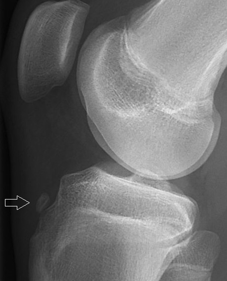
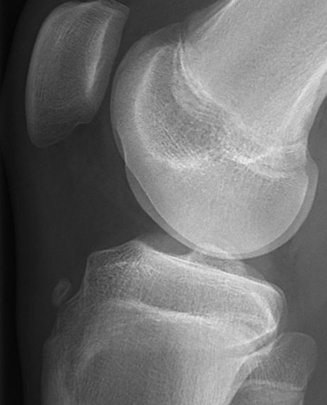
Avulsion fractures can occur in various parts of the body. Dental avulsion is the complete displacement of a tooth from its socket, requiring prompt management within 20–40 minutes to improve prognosis. The tuberosity avulsion fracture of the fifth metatarsal, also called pseudo-Jones or dancer's fracture, is commonly caused by the lateral band of the plantar aponeurosis and typically heals within eight weeks with a hard-soled shoe or walking cast. Tibial tuberosity avulsion fractures result from violent quadriceps contraction, often from high-power jumps, and are most common in teenagers active in sports, with a link to Osgood-Schlatter's disease.
Reader's Guide
Avulsion fractures represent a distinct category of bone injury where a fragment is pulled away rather than the bone breaking across its shaft. Their significance lies in the mechanism—often involving tendon or ligament attachment—and the specific treatment required. Small avulsions may heal with rest and support, but displaced fractures often need surgical intervention such as open reduction and internal fixation. The condition highlights the interplay between muscle strength and bone integrity, especially in trained athletes who can overcome neurological limits. In paleontology, evidence of tendon avulsions in theropod dinosaurs like Tyrannosaurus and Allosaurus suggests complex musculature different from birds. Understanding avulsion fractures aids in proper diagnosis and management, preventing long-term disability.
Did You Know?
- Dental avulsion requires management within 20–40 minutes of injury for best prognosis.
- Tuberosity avulsion of the 5th metatarsal is also known as pseudo-Jones fracture or dancer's fracture.
- Tibial tuberosity avulsion fractures are most common in teenagers and linked to Osgood-Schlatter's disease.
- Avulsion injuries in dinosaurs have been documented in multiple genera (e.g., Tyrannosaurus, Allosaurus, Acrocanthosaurus, Deinonychus) and on various bones, not exclusively on the humerus and scapula.
Mechanism and the Role of Neurological Inhibition
An avulsion fracture occurs when a small fragment of bone is ripped away from the main bone mass due to physical trauma. The mechanism can take two distinct paths: at a ligament, where external forces like a fall or a sudden pull yank the attachment point, or at a tendon, where an internal muscular contraction generates enough force to overcome the structural integrity of the bone itself. Under normal circumstances, the human nervous system acts as a built-in safety mechanism, placing neurological limits on how forcefully a muscle can contract. This inhibition prevents the muscle from generating enough power to tear a bone fragment free. However, highly trained athletes can push past these neurological brakes, producing a significantly greater force output. This enhanced capability, while advantageous for performance, also elevates the risk of avulsing a bone fragment—making the very training that builds elite strength a potential vulnerability.
Clinical Subtypes and Their Management
Avulsion fractures manifest in several well-recognized clinical forms, each with its own etiology and recovery pathway. Dental avulsion involves the complete traumatic displacement of a tooth from its alveolar socket and is treated as a serious dental emergency; the prognosis for saving the tooth hinges on receiving prompt management within a narrow 20-to-40-minute window after the injury. The tuberosity avulsion of the fifth metatarsal—sometimes called a pseudo-Jones or dancer's fracture—arises most likely from the lateral band of the plantar aponeurosis and is typically managed with a hard-soled shoe or walking cast until pain resolves, with healing generally completing within eight weeks. Tibial tuberosity avulsion, by contrast, results from a violent quadriceps contraction, often during a high-power jump, and strikes teenagers who are heavily involved in sport. Incomplete cases respond to the RICE protocol, while complete or displaced fractures usually demand surgical pinning. Research has also linked a prior history of Osgood-Schlatter's disease to increased susceptibility.
Paleontological Evidence in Theropod Dinosaurs
In 2001, paleontologist Bruce Rothschild and colleagues published a study that brought avulsion fractures into the prehistoric record. By examining skeletal remains of theropod dinosaurs, the team identified tendon avulsion scars exclusively in two genera: Tyrannosaurus and Allosaurus. These injury marks were confined to the humerus and scapula, suggesting that the affected muscles anchored at those specific sites. One particularly notable example is a divot found on the humerus of Sue, the famous T. rex specimen. The location of this divot corresponds to the origin of either the deltoid or teres major muscles. The researchers drew a broader anatomical inference from these findings: the pattern of avulsion scarring on theropod scapulae implies that these dinosaurs possessed a musculature that was more complex and functionally distinct from that of their living descendants, the birds. In other words, the very injuries preserved in fossil bone offer a window into how theropods moved and powered their limbs.
Treatment Modalities and Surgical Intervention
The management of an avulsion fracture is dictated largely by the size and displacement of the torn fragment. When the fracture is small and the bone piece remains in place, conservative measures are typically sufficient: rest, a support bandage, and the application of ice to reduce swelling. The patient can often resume normal activities once pain subsides, as seen in the typical eight-week healing trajectory of a fifth metatarsal tuberosity avulsion. However, when the fragment is displaced, surgical intervention becomes necessary. Two principal approaches exist. Open reduction and internal fixation involves making a surgical incision to directly visualize the fracture site, then securing the bone fragment with pins, screws, or similar hardware. Closed reduction and pinning, by contrast, achieves realignment without a full open incision, guiding the fragment back into position and stabilizing it with percutaneous pins. The choice between these techniques depends on the degree of displacement and the specific anatomical site involved, with complete tibial tuberosity avulsions being a classic indication for surgical pinning.
Gallery
Frequently Asked Questions
Who is Avulsion fracture?
Avulsion fracture is a subtype of traumatic bone break in which a small chip is torn free from the main bone mass, usually because a ligament or tendon exerts more pulling force than the attachment point can endure. It sits in the broader family of stress-and-trauma fractures but is defined specifically by that 'piece ripped away' mechanism.
What are Avulsion fracture's powers/role?
Its signature 'ability' is to detach a bone fragment when either an external force (like a fall that yanks a ligament) or an internal muscular contraction overcomes the structural bond at the insertion site. Contrary to older textbook simplifications, this mechanism is not restricted to elite athletes; it can strike anyone whose muscle or ligament load briefly exceeds bone-holding strength.
Where does Avulsion fracture most often appear?
The three sites most frequently cited in the medical canon are the dental socket, the tuberosity of the fifth metatarsal, and the tibial tuberosity. All three are anchor points where tendons or ligaments grip bone, making them natural weak spots for the pulling action that defines this fracture type.
How does Avulsion fracture's story end (treatment)?
For non-displaced cases, the typical 'resolution arc' is conservative: rest, a supportive bandage, and ice to control swelling while the fragment heals in place. When the torn piece has shifted out of alignment, surgery is required to reposition or fix the fragment so the bone can knit back together properly.
Why is Avulsion fracture important in the dinosaur fossil record?
Avulsion fractures have been identified in the skeletal remains of several large theropods, including Tyrannosaurus, Allosaurus, Acrocanthosaurus, and Deinonychus. Their presence in these fossils shows that even massive prehistoric predators were subject to the same tendon- and ligament-pulling mechanics that produce this fracture in modern patients.
More in Bone fractures 1-24
Spotted an error? Know more?
This is a living reference — every entry is fact-audited, and reader corrections feed straight into our audit queue. Suggest an edit · See this site's audit record