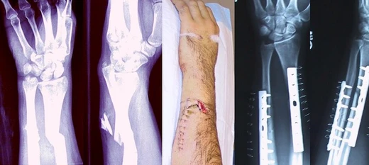
Bosworth fracture
Rare ankle fracture with fixed posterior fibular dislocation.

The Bosworth fracture is an uncommon ankle injury involving a break in the lower fibula, where the upper part of the broken bone becomes locked behind a bony ridge on the back of the tibia. This dislocation is fixed, meaning the fragment is stuck in place. The injury results from a forceful outward twisting of the ankle, and the foot remains twisted outward afterward. This persistent rotation can make X-rays hard to read, often leading to a wrong diagnosis and improper care.
Because the fibular fragment is wedged behind the tibia, closed reduction (manipulating the bone without surgery) usually fails. Therefore, early open surgery to realign and fix the bones is standard to prevent additional problems.
Compared to other ankle fracture-dislocations, the Bosworth injury carries a higher risk of complications. These include skin death, surface infection, increased pressure within the leg (compartment syndrome), death of the ankle bone (astragalus avascular necrosis), joint stiffness, nerve damage, and later arthritis. Delaying surgery or repeatedly attempting closed reduction raises the chance of poor outcomes and more complications.
- field
- Orthopedic surgery
- known_for
- Description of the Bosworth fracture
- first_described_by
- David M. Bosworth in 1947
- mechanism
- Severe external rotation of the ankle
- treatment
- Open reduction internal fixation
Lore & Background
The entrapment of an intact fibula behind the tibia was described by Ashhurst and Bromer in 1922, who attributed the description of the mechanism of injury to Huguier's 1848 publication. The injury involving fibular fracture with posterior dislocation was described by David M. Bosworth in 1947. Because of a fixed dislocation within the proximal fibular fragment posterior of the lateral ridge of the tibia, the Bosworth fracture is typically irreducible using closed techniques, requiring early open reduction to avoid further complications.
Reader's Guide
The Bosworth fracture is significant as a distinct and often misdiagnosed ankle injury due to the persistent external rotation of the ankle, which complicates radiographic interpretation. Its rarity and the fixed posterior dislocation of the fibular fragment behind the tibia make closed reduction difficult, necessitating open reduction internal fixation. Delays in surgical reduction or repeated closed reduction efforts increase the risk of complications such as cutaneous necrosis, superficial infection, compartmental syndrome, astragalus avascular necrosis, joint stiffness, superficial fibular nerve lesion, and secondary arthrosis. The injury's description by David M. Bosworth in 1947 built upon earlier observations by Ashhurst and Bromer and Huguier, highlighting the evolution of orthopedic understanding of ankle trauma. Its legacy lies in emphasizing the need for prompt, accurate diagnosis and surgical intervention to prevent poor outcomes.
Did You Know?
- The Bosworth fracture involves a fixed posterior dislocation of the proximal fibular fragment behind the posterior tibial tubercle.
- The injury is caused by severe external rotation of the ankle, and the ankle remains externally rotated after injury.
- Closed reduction is typically impossible due to entrapment of the fibula behind the tibia.
- Complications are more common in Bosworth fractures than in any other type of ankle fracture-dislocation.
Why a Broken Bone Hurts
Although bone tissue itself is devoid of pain-sensing nerve endings, a fracture is nonetheless an intensely painful event. The discomfort originates from several sources beyond the bone shaft. The periosteum and, in some cases, the endosteum—both richly threaded with nociceptors—suffer a break in continuity when the bone snaps, sending sharp signals to the brain. Ruptured marrow spills blood into surrounding soft tissue, creating edema and a hematoma that press against nearby structures and generate a deep, throbbing ache. The body's reflexive response compounds the suffering: surrounding muscles lock into involuntary spasms, attempting to immobilize the displaced fragments. In more severe presentations, the injury extends beyond the bone itself. Nerves, blood vessels, and muscles in the vicinity may be lacerated or compressed. Spinal fractures can impinge on the spinal cord or nerve roots, while cranial fractures threaten the contents of the skull, each producing its own constellation of neurological signs that demand urgent medical evaluation to prevent cascading damage.
The Body's Repair Blueprint
The healing of a fractured bone unfolds in a carefully sequenced cascade. Immediately after the break, bleeding from the injured bone and adjacent tissues pools between the fragments, forming a fracture hematoma. This blood coagulates, creating a provisional scaffold. Over the following days, new capillaries invade the gelatinous clot, delivering phagocytes that clear away dead cellular debris. The same vessels carry fibroblasts, which proliferate and secrete collagen fibers, gradually replacing the clot with a flexible collagen matrix. This rubbery bridge permits only minimal movement of the fragments unless subjected to extreme force. Progressively, certain fibroblasts begin depositing bone matrix as collagen monomers that spontaneously assemble, with calcium hydroxyapatite crystals locking in as insoluble mineral deposits. This mineralization transforms the soft matrix into rigid bone. The resulting woven bone, visible on X-ray within roughly six weeks in adults and sooner in children, lacks the mechanical strength of mature tissue. Through a prolonged remodelling phase, it is exchanged for organized lamellar bone—a process that can stretch to eighteen months. In adults, functional strength typically reaches about eighty percent of normal by three months. Adequate nutrition and calcium intake support this trajectory, while tobacco use measurably impairs it.
When Healing Goes Sideways
Not every fracture follows a smooth recovery path. Among the most feared sequelae is compartment syndrome, a condition in which pressure within a muscle compartment escalates to the point of threatening tissue viability; left untreated, it can ultimately necessitate amputation of the affected limb. Two other healing failures carry distinct names: non-union, in which the fractured bone simply fails to knit back together, and malunion, in which it does heal but in a deformed configuration. A particularly troublesome variant of malunion is malrotation, where the bone sets at an incorrect angle around its long axis—a problem especially common after fractures of the femur or tibia. Clinicians organize these complications along a temporal axis into three broad categories. Immediate complications manifest at the very moment of injury. Early complications emerge during the first few days following the fracture. Late complications surface weeks, months, or even longer after the initial event. This time-based framework helps clinicians anticipate which risks are most pressing at each stage of care and tailor surveillance accordingly, ensuring that a seemingly stable fracture does not quietly evolve into a far more dangerous problem.
Naming, Sorting, and Confirming
In orthopedic practice, fractures are sorted using multiple overlapping schemes. Historically, many fracture patterns bear the name of the physician who first described them, though modern classification systems add greater systematic rigor. A fundamental distinction separates stable fractures—those unlikely to shift further—from unstable ones. By mechanism, a break may be traumatic (resulting from a fall, collision, or direct impact), pathologic (occurring through a bone already weakened by disease such as osteoporosis, bone cancer, or osteogenesis imperfecta), or periprosthetic (arising at the mechanical stress point adjacent to an implanted device). Soft-tissue integrity provides another axis: closed or simple fractures leave the overlying skin intact, whereas open or compound fractures create a wound that communicates with the fracture site, exposing bone to contamination. The infection risk after internal fixation jumps from one to two percent in closed fractures to as high as thirty percent in open ones. Displacement patterns—non-displaced, translated, angulated, rotated, or shortened—further refine the description. Diagnosis typically begins with history and physical examination, confirmed by radiographic imaging, with CT or MRI reserved for cases where standard X-rays prove insufficient.
Frequently Asked Questions
What is a Bosworth fracture?
It is a rare ankle injury in which the lower fibula snaps and the upper fragment wedges itself behind a bony ridge on the posterior tibia, creating a fixed dislocation that cannot be popped back into place by hand.
Who is the Bosworth fracture named after?
Orthopedic surgeon David M. Bosworth first described this specific pattern of ankle injury in 1947, and the eponym stuck in the literature ever since.
What mechanism produces a Bosworth fracture?
A violent external-rotation force on the ankle is what drives the fibular fragment behind the tibia, and the foot typically remains twisted outward after the event.
Why do doctors often miss a Bosworth fracture on imaging?
Because the foot stays locked in that outward rotation, standard X-ray angles become distorted and hard to read, which routinely leads to a misdiagnosis and a course of treatment that doesn't address the true injury.
How is a Bosworth fracture treated?
Closed reduction—manipulating the bones back without cutting—generally fails because the fragment is mechanically trapped behind the tibia, so the standard of care is open reduction with internal fixation to free and stabilize the bone.
More in Bone fractures 1-24
Spotted an error? Know more?
This is a living reference — every entry is fact-audited, and reader corrections feed straight into our audit queue. Suggest an edit · See this site's audit record